A CHIVA Strategy That Departs from the Classical Staging Sequence: Clinical Results and Implications — Less Is More
Key Takeaways
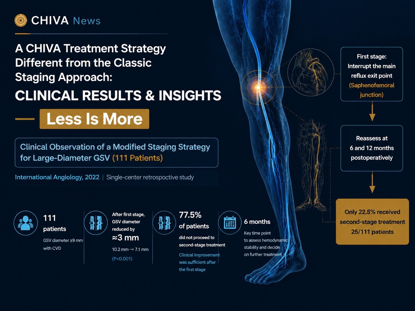
In 2022, a team from Vall d’Hebron University Hospital in Barcelona, Spain published a retrospective observational study in International Angiology on CHIVA treatment of large-diameter great saphenous veins. The study included 111 patients with chronic venous disease and a great saphenous vein (GSV) diameter of 9 mm or greater, treated between January 2010 and December 2019. A CHIVA staging strategy modified for large-diameter GSVs was used, and the authors followed changes in GSV diameter after the first stage, clinical improvement, the subsequent need for second-stage treatment, and related complications.
Within this cohort, 79.3% had a Type III shunt (hereafter Shunt III) and 20.7% had a Type I+II shunt. The study therefore provides not only clinical data on large-diameter GSVs, but also a body of CHIVA observations in which large-diameter Shunt III predominates.
A design that differs from the classical CHIVA staging sequence
Shunt III in CHIVA commonly involves staged treatment. The authors note that in large-diameter GSVs the risk of complications is higher when the first and second stages are performed in the same session, and that they therefore adopted a modified two-stage strategy whose immediate aim was to reduce the rate of symptomatic superficial vein thrombosis after treatment.
In their discussion, the authors point out that the classical two-stage approach to Shunt III usually addresses the incompetent saphenous tributaries first. This study reversed that order: the first stage interrupted the main escape point — in every patient in this cohort, an incompetent saphenofemoral junction — after which clinical improvement and duplex follow-up determined whether a second stage was needed at all.
The purpose of the first stage was to lower pressure and flow load within the GSV by interrupting the main source of reflux, thereby reducing vein diameter. Only if clinical improvement remained insufficient would the incompetent N3/N4 tributaries be addressed. The second stage was not a predetermined step. Patients were reassessed at 6 and 12 months after the procedure, and second-stage treatment was performed only in those with persistent symptoms or insufficient clinical improvement whose GSV had also shrunk to a diameter suitable for further treatment.
What this staging modifies, then, is not only the surgical sequence but the temporal logic of the treatment decision: complete the key hemodynamic correction first, and let the resulting state determine the next step.
After the first stage, the great saphenous vein narrowed by roughly 3 mm on average
All 111 patients underwent the first stage. After interruption of the main escape point, mean GSV diameter fell from 10.2 ± 1.1 mm before treatment to 7.1 ± 1.5 mm, a mean reduction of 3 ± 1.6 mm, a statistically significant difference (P < 0.001). The larger the baseline GSV diameter, the greater the post-treatment reduction.
The authors also observed that most of the change in GSV diameter occurred within the first 6 months and stabilized thereafter. On this basis they consider approximately 6 months to be an important point at which to judge whether the hemodynamic state has stabilized and whether a second stage is required.
This finding also gives “large diameter” a more dynamic meaning.
Vein diameter is not a fixed anatomical measure existing independently of hemodynamic conditions. It is influenced by intravenous pressure, flow load, and drainage conditions. Once the principal source of abnormal flow has been corrected, a preserved great saphenous vein can retract appreciably as its hemodynamic environment changes.
In this cohort, that change was not confined to imaging: it went on to shape the subsequent treatment pathway.
77.5% of patients did not proceed to a second stage
Of the 111 patients, only 25 (22.5%) ultimately received second-stage treatment. The remaining 86 patients — 77.5% — had achieved sufficient clinical improvement after first-stage interruption of the saphenofemoral junction and underwent no further surgery.
CEAP clinical class also improved significantly after the first stage (P < 0.001).
The first stage therefore altered GSV diameter, the patient’s clinical status, and the subsequent need for treatment at the same time. N3/N4 tributaries that might have been considered for further treatment on the basis of the original, pre-treatment hemodynamic picture did not necessarily still require the same intervention once the principal abnormal load had been corrected.
This is a defining feature of the staging strategy in this study: the second stage is not a prescribed step that follows the first, but a fresh decision triggered by what the first stage achieved.
For 77.5% of patients, the improvement after the first stage was sufficient, and treatment stopped there.
Implications for large-diameter Shunt III
Eighty-eight of the 111 patients had Shunt III, or 79.3%. This composition makes the study particularly relevant to large-diameter Shunt III.
Classical Shunt III staging rests on a well-established hemodynamic logic. This study offers an alternative sequence: under the specific condition of a markedly dilated GSV, interrupt the main abnormal N1→N2 connection first, observe how N2 and its associated N3 network behave under the new pressure and drainage conditions, and only then decide whether N2→N3 still requires treatment.
The findings do not support the conclusion that all Shunt III cases should depart from the classical sequence, but they do suggest that under the specific condition of a large diameter, the treatment sequence itself can become part of an individualized hemodynamic strategy.
Once the principal source of abnormal pressure is brought under control, GSV diameter, the load on the superficial network, clinical presentation, and the patient’s subsequent treatment needs may all change. The hemodynamic configuration seen before treatment is the starting point for planning; the new state that emerges after the first stage can equally serve as the basis for planning what comes next.
For large-diameter Shunt III, this produces a dynamic clinical pathway: correct the key abnormal flow, observe how the venous system re-establishes balance, and then decide whether further intervention is still needed.
Large diameter as a strategic parameter, not a simple treatment threshold
The authors selected patients with a GSV diameter of 9 mm or greater not because 9 mm defines whether CHIVA can be performed or whether the GSV can be preserved, but because large diameter itself is associated with a higher rate of symptomatic superficial vein thrombosis after treatment.
Previous work cited in the paper indicates that when GSV diameter exceeds 8.5 mm, the relative risk of symptomatic superficial vein thrombosis (SVT) after treatment is approximately 3.1 times that seen at diameters of 8.5 mm or less.
Symptomatic SVT is not unique to CHIVA. The authors note that in patients with large-diameter GSVs, symptomatic SVT may likewise occur after sclerotherapy, radiofrequency ablation, and endovenous laser treatment. The mechanisms by which it develops, however, are not identical across these modalities.
In their review of the characteristics and results of CHIVA, Faccini, Ermini, and Franceschi discuss post-CHIVA GSV thrombophlebitis specifically. They state that CHIVA does not aim to damage or occlude the great saphenous vein, so the procedure does not injure the endothelium along the whole length of the vessel; in their account, GSV thrombophlebitis after CHIVA relates to the temporary loss of the previous centrifugal flow following the hemodynamic change and to the resulting local low-flow state, which differs from the thrombotic background created by foam sclerotherapy or endovenous ablation through venous wall and endothelial injury and deliberate occlusion of the target vein.
Once an effective new re-entry point is established, GSV flow can be restored, and this recanalization and recovery typically occurs within a few months of the procedure. Within the hemodynamic framework of CHIVA, therefore, such events cannot simply be equated with the permanent occlusion of a target vein in other techniques, still less read as treatment failure.
This distinction connects naturally to the staging design used here. In large-diameter GSVs, which already carry a higher rate of SVT, the authors first interrupted the main source of reflux, lowering pressure and flow within the GSV. After the first stage, mean GSV diameter fell from 10.2 mm to approximately 7.1 mm; only patients whose clinical improvement remained insufficient went on to second-stage N3/N4 treatment under these new flow conditions.
Sixteen patients in the study developed SVT, an overall rate of 14.4% (16/111); of these, 12 were symptomatic, for a symptomatic SVT rate of 10.8% (12/111). Patients who developed SVT also had a larger mean pre-treatment GSV diameter.
The staging design is thus not a response to some hazard peculiar to CHIVA. It is an attempt, through a change in treatment sequence, to make the hemodynamic transition more gradual, to further reduce the rate of symptomatic SVT after treatment, and to preserve the venous drainage network as completely as possible.
Put differently, the authors staged treatment not because a large-diameter vein cannot be preserved, but precisely in order to optimize the treatment process while continuing to preserve the GSV.
Seen this way, large diameter is better understood as a strategic parameter: it can inform risk assessment and guide the design of individualized treatment plans, the timing of follow-up, and the form of any subsequent intervention.
Less Is More: the clinical significance behind the staging strategy
CHIVA is a treatment concept built on a strong commitment to individualized planning. It calls for understanding the characteristics of the venous-venous shunt, following the principle of preserving the superficial venous drainage network and its function as far as possible, and designing an individualized strategy around the patient’s particular circumstances and treatment goals. The strategy in this study rests on a dynamic clinical pathway: complete the key hemodynamic correction first, then let the clinical and duplex changes that actually occur after the first stage determine whether further treatment remains necessary.
Close to 80% of patients in this study never proceeded to a second stage. Under this staging strategy, most patients not only saw a marked reduction in GSV diameter after the first stage but — more importantly — achieved sufficient clinical improvement, so that the N3/N4 network that might otherwise have been treated no longer required intervention.
From the standpoint of the clinical pathway, this means some patients can avoid a second procedure and the burden that comes with it. From the standpoint of vein preservation, it means the superficial venous network is treated further only where a clear need remains, leaving more room to preserve veins that still carry effective drainage value.
This staging model also extends what “individualization” means in CHIVA. Individualization is not only a matter of designing different plans for different shunt configurations; it also means that the plan for a single patient can continue to be adjusted as the hemodynamic state changes after treatment. The first-stage plan derives from the pre-treatment hemodynamic assessment; whether a second stage follows derives from the clinical picture and the new hemodynamic state after the first.
In large-diameter Shunt III particularly, this study offers a strategy of real practical value: the classical principles still form the basis of treatment design, but under specific hemodynamic conditions the staging sequence and the eventual scope of intervention can be individualized further. The goal is not to complete every step planned before treatment, but to correct the principal abnormal pressure and shunt through the interventions that are required, and then — once clinical and hemodynamic improvement is sufficient — to decide whether anything further is needed.
The value this study brings to clinical practice, therefore, is not simply a result of “a mean 3 mm reduction in GSV diameter,” nor merely the technical question of how large-diameter GSVs can be managed. It suggests something further: that the treatment sequence itself can be part of a hemodynamic strategy, and that the response to treatment should be part of the next decision.
Reducing interventions that are no longer necessary through more targeted staging, while maintaining clinical effectiveness, improving the patient’s whole course of treatment and recovery, and preserving valuable veins and their drainage network wherever possible — this may be what most deserves attention in the study for clinical practice.
Less Is More, here, does not mean doing less than is necessary. It means that every step of intervention has a clear hemodynamic justification, and that when treatment has been enough, one knows when to stop.
Study limitations
This is a single-center, retrospective observational study with no concurrent control group. The authors note that case selection, missing data in some records, and sample size may all have influenced the results, and consider that larger prospective studies are still needed to validate this strategy in patients with large-diameter GSVs.
The available data therefore cannot establish that all large-diameter GSVs or all Shunt III cases should follow the same staging sequence, nor can they show this strategy to be superior to other CHIVA pathways or to other venous treatments.
As a clinical study in which large-diameter Shunt III predominates, however, it points to a direction worth validating further: beneath the established hemodynamic principles of CHIVA, there is still room to individualize and refine treatment sequence, treatment timing, and the eventual scope of intervention.
For the clinician, what this study finally leaves is not a fixed rule for how a vein of 9 mm or more should be treated, but a way of deciding that is characteristically CHIVA: correct the key hemodynamic abnormality, observe how the venous system re-establishes balance, and then determine what still needs to be done — and what no longer does.
References
Tenezaca-Sari X, García-Reyes M, Escribano-Ferrer JM, Marrero C, Bellmunt-Montoya S. The CHIVA strategy applied to large-diameter saphenous veins. Int Angiol. 2022;41(4):332-337. DOI: 10.23736/S0392-9590.22.04831-3
Faccini FP, Ermini S, Franceschi C. CHIVA to treat saphenous vein insufficiency in chronic venous disease: characteristics and results. J Vasc Bras. 2019;18:e20180099.
About the Authors
This study was conducted by the Department of Angiology, Vascular and Endovascular Surgery at Vall d’Hebron University Hospital in Barcelona, Spain. The first author is Xavier Tenezaca-Sari. It was published in International Angiology.
Disclaimer
This article is compiled from publicly published literature for professional information exchange and content research. It does not constitute specific medical advice.