
From a CHIVA Perspective, What Is Still Missing After the UK’s New Venous Ultrasound Protocol Standardizes Mapping?
From a CHIVA Perspective, What Is Still Missing After the UK’s New Venous Ultrasound Protocol Standardizes Mapping?
Key Points
Study basis:
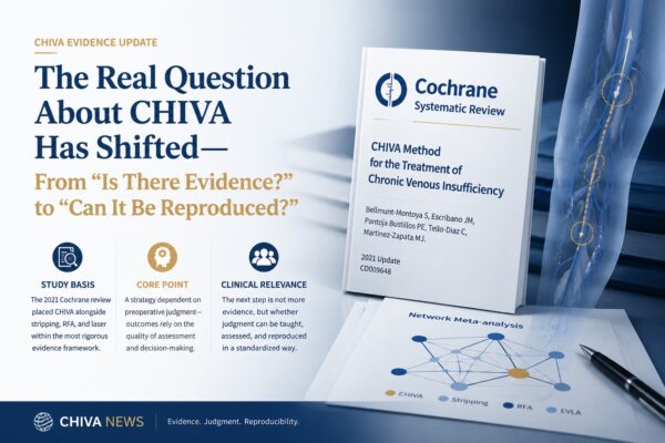
In April 2026, The College and Society for Clinical Vascular Science (CSVS) in the UK released the Lower Limb Venous Duplex Ultrasound Examination Scan Protocol, Version 2.0 (Doc Ref PS-PG004), updating the 1.0 version published in April 2021. The protocol was prepared by the CSVS Professional Standards Committee, approved by the Executive Committee, and is scheduled for review in April 2028.
Core argument:
CSVS Version 2.0 incorporates key actions such as standing assessment, unified reflux time definitions, complete venous mapping, and reflux source tracing into a standardized workflow. Compared with many countries and regions that still lack a unified lower limb venous ultrasound protocol, this document provides a clearer methodological baseline for venous reflux assessment.
Clinical implication:
From a CHIVA perspective, the progress of the protocol deserves recognition, but so do its boundaries. It standardizes how to examine, describe, and trace reflux, but it does not interpret the functional significance of reflux structures, nor does it enter the level of shunt classification, procedure selection, or treatment decision-making. After mapping has been standardized, a further methodological system is still needed to convert an ultrasound map into a hemodynamic treatment strategy.
CHIVA is a treatment strategy that relies heavily on preoperative ultrasound judgment. Every ligation decision is based on prior understanding of the individual patient’s flow distribution, reflux source, drainage pathway, and compensatory channels.
For that reason, CHIVA requires more from ultrasound than simply “detecting reflux.” It requires the operator to read treatment strategy from the reflux map. This is exactly why CSVS Version 2.0 is worth re-examining from a CHIVA perspective: it moves generic diagnostic workflow one step forward, but it still preserves a clear boundary at the level of treatment decision-making.
1. Standing assessment has been further standardized
The protocol clearly states that the supine position is not appropriate for evaluating lower limb venous reflux, and that patients should be examined in the standing position, or as close to standing as possible.
This requirement is not new in hemodynamic terms, but the fact that it has been written into a professional scanning protocol means that a key examination principle has been converted into a standard action that can be taught, performed, and audited. For regions where clinical practice is still inconsistent, that alone represents meaningful progress.
For CHIVA, standing assessment is a basic condition. The identification of escape points, reflux pathways, shunt structures, and re-entry depends on real flow patterns under the effect of gravity. If the positional condition is not valid, subsequent interpretation may already be compromised at its source.
2. Reflux must be defined consistently before findings can be comparable
The protocol applies explicit time thresholds for superficial venous reflux and deep venous reflux. The values themselves are part of widely accepted international standards. What matters more is that they are placed within a unified interpretive framework.
Across centers, consistency in reflux definition is fundamental. If operators do not use the same standard for deciding what counts as reflux, then case discussion, treatment selection, and outcome comparison lose a common language.
From a CHIVA perspective, this matters just as much. The choice between CHIVA 1, CHIVA 2, or CHIVA 1+2 must be based on clear assessment of reflux status in different venous segments. Only when reflux definitions are relatively consistent can shunt analysis begin to achieve cross-center comparability.
At the same time, reflux time thresholds remain only a classification tool. They can help determine whether a venous segment has reflux, but they cannot explain the functional role of that reflux within the overall drainage network.
3. Complete mapping raises the lower limit of examination quality
CSVS Version 2.0 gives vein mapping its own dedicated importance and requires that varicose veins that cannot be simply assigned to the great saphenous or small saphenous systems should be traced upward along the reflux path to identify their source, while also covering systematic assessment of medial, anterior, lateral, and posterior regions.
This point deserves particular recognition. It means venous ultrasound should not stop at limited segmental scanning, nor merely describe an enlarged vessel segment. It should go as far as possible toward reconstructing the source and course of reflux. Compared with clinical environments where complete mapping is not routine, this clearly raises the lower limit of examination quality.
This is also highly relevant to the hemodynamic mapping long emphasized by CHIVA. CHIVA is not about identifying which vein looks enlarged, nor simply detecting which segment has reflux. It is about understanding pressure transmission and drainage relationships across the superficial venous network.
But mapping itself is still a map, not a treatment plan.
A general ultrasound protocol can require the operator to trace the source of reflux, but it will not go on to determine how that source should be managed in treatment. It can require a complete venous map, but it will not indicate which channels should be preserved, which connection points should be interrupted, or which apparently abnormal flows are in fact serving useful drainage or compensatory functions.
4. From a CHIVA perspective, the limitation lies not in the protocol itself, but in its level
CSVS Version 2.0 is a lower limb venous duplex ultrasound examination protocol. Its function is to standardize examination workflow and the basis of reporting, not to prescribe the execution pathway of any particular treatment method.
It therefore does not address shunt classification, does not address the choice between CHIVA 1, CHIVA 2, or CHIVA 1+2, and does not discuss how to reconstruct a more rational hemodynamic order through limited intervention while preserving venous resources.
This is not a flaw in the protocol. It is the boundary of its function.
In traditional varicose vein treatment, mapping often serves a relatively direct aim: identifying which diseased veins need to be ablated, stripped, closed, or otherwise treated. In that context, the ultrasound map functions mainly as a lesion-localization map.
CHIVA uses mapping differently. It must determine not only where reflux exists, but whether that reflux constitutes a pathological entry point, whether an effective drainage outlet is present, whether the channel has preservation value, and whether limited intervention can change the overall pressure distribution.
5. After mapping has been standardized, the real challenge begins
CSVS Version 2.0 makes several key components of lower limb venous reflux examination clearer: standing assessment, unified reflux definition, complete mapping, and reflux source tracing. These provide a necessary foundation for higher-quality venous diagnosis and treatment.
But from a CHIVA perspective, diagnostic standardization is not the same as treatment strategy standardization.
Once a venous map has been drawn, the real questions begin: which reflux pathways need to be interrupted? Which channels should be preserved? Which dilated veins are merely consequences rather than pressure sources? Which drainage outlets, if closed, may actually damage existing compensation?
These questions cannot be answered by a general ultrasound protocol alone. They require CHIVA’s own hemodynamic interpretive framework, its logic of procedure selection, and execution standards that can be trained, assessed, and reproduced.
In this sense, the value of the UK’s new venous ultrasound protocol lies in further standardizing the diagnostic foundation required for modern venous treatment. What CHIVA still needs to accomplish is the critical translation from mapping to treatment decision-making. This is precisely the level of standardization that the Global CHIVA Program is now facing.
For CHIVA to enter broader clinical practice, it needs both high-quality ultrasound foundations and a treatment decision system that can be taught, evaluated, and reproduced. CSVS Version 2.0 shows that venous care is placing increasing emphasis on standardized mapping. From a CHIVA perspective, however, the real challenge begins exactly after mapping is completed.
Reference Source
The College and Society for Clinical Vascular Science (CSVS). Lower Limb Venous Duplex Ultrasound Examination Scan Protocol. Version 2.0, April 2026, Doc Ref PS-PG004. Review April 2028.
About the Publishing Body
The College and Society for Clinical Vascular Science (CSVS), founded in 1992, is one of the key UK bodies responsible for professional standards, accreditation, and continuing education in clinical vascular science. Its Vascular Science Guidelines series is intended for the UK and the broader vascular science community, serving as an important methodological reference for non-invasive vascular diagnostic services. CSVS explicitly states that these documents do not replace clinical judgment or local pathways, but they do represent a methodological baseline within UK vascular ultrasound practice.
Note
This article is based on publicly available literature and documents, and is intended for professional information exchange and content research only. It does not constitute specific medical advice.